

Recognizing Signs That You Need Emergency Dentistry
February 26, 2026
Pregnancy Support Systems and Resources
February 26, 2026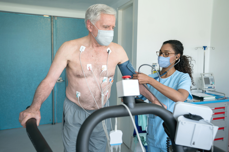
Your upcoming nuclear stress test will provide data about your heart’s health. A nuclear stress test evaluates blood flow and it identifies areas with poor circulation. Because your heart requires oxygen, the test measures supply and assesses function. This procedure is a standard tool for assessing risk.
Tracer Injection Mechanics
The nuclear stress test begins with a small injection of a radioactive tracer into a vein. This substance travels through the blood, and heart muscle cells absorb the material. The level of tracer uptake in heart tissue depends on the amount of blood that reaches each area. When the tracer reaches the heart, it emits energy, and a camera detects it. Tissues with good blood supply absorb the tracer well, so those areas appear bright. Regions with reduced blood flow take up less tracer and look darker. This material is not a dye, but it acts like a molecular beacon. The absorption difference helps identify parts of your heart muscle that are not receiving enough oxygen-rich blood. If some areas absorb less tracer, the doctor will examine these regions further. The visual separation of healthy and at-risk tissue gives the healthcare team information about your heart.
Stress Induction Process
If you are able, you will perform physical activity to increase the heart’s demand for blood. This phase is an exercise stress test, and you walk on a treadmill. If you cannot exercise, the doctor may prescribe medication that mimics the effects of running. During the test, your heart rate and blood pressure rise. This mimics vigorous daily activity. When the heart beats faster during stress, it needs more oxygen. The coronary arteries expand to supply extra blood. The test is carefully monitored, and the healthcare team observes how your heart responds to increased workload. As your heart works harder, the medical team carefully watches the tracer’s distribution.
Areas receiving enough oxygenated blood will show bright signals, while segments that do not keep up with demand appear darker. They compare this stress state to rest, and look for significant differences. These changes help detect blockages or narrowed arteries. This comparison helps reveal issues like angina and heart attack risks that appear only under exertion. In people with a history of arrhythmias or those at risk, the test sometimes reveals abnormal rhythms that require further attention. By identifying these concerns, your doctor can recommend strategies or treatments.
Image Capture Technology
A specialized gamma camera rotates around your chest. It captures the energy emitted by the tracer in your heart tissue. This camera is sensitive to low levels of radiation, and it works with detectors that produce cross-sectional images. After image capture, computer software processes and reconstructs these images. This lets your healthcare team see your heart’s structure and blood flow clearly. The software highlights potential blockages and compares images taken during rest and stress phases. This process helps the medical team spot problems such as narrowed arteries, weakened heart muscle, or damage from a previous heart attack. Dark spots on the scan indicate areas where blood flow is restricted or absent.
Nuclear Stress Test Preparation
You play an active role in the accuracy of these scientific results. Please wear comfortable clothing, and bring a list of your current medications. Adhering to these guidelines gives clear images and prevents the need for retesting. Contact your office today if you have questions about your instructions.


