

The Intersection of Pediatric Neurology and Mental Health
October 4, 2025
How Emergency Rooms Handle Trauma Cases
October 6, 2025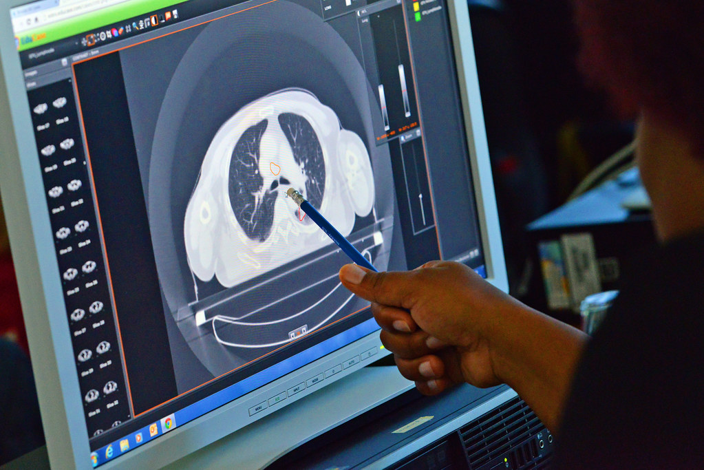
Computed tomography (CT) scans use advanced X-ray technology to produce detailed three-dimensional images of bones and soft tissues. These scans help physicians diagnose various conditions and inform treatment decisions. They play a key role in modern imaging by providing clear, accurate internal views. Here are a few things to know about the myths and facts about CT scans and radiation exposure:
Understanding a CT Scan
A CT scan is a specialized form of X-ray imaging. While it captures images of bones like standard X-rays, it also employs advanced technology to create three-dimensional pictures. These 3D images provide views of organs and other soft tissues, offering more detailed information than conventional X-rays.
The detailed imaging from a CT scan helps physicians assess a wide range of health conditions. They can examine internal injuries, fractures, and head trauma, as well as identify spine disease, tumors, and arterial issues. Additionally, some doctors utilize scans to monitor the progress of ongoing treatments. This combination of diagnostic and monitoring capabilities makes CT scans a versatile tool in patient care.
How a Scan Works
The machine features a large, circular opening through which a narrow X-ray beam rotates around your body, creating cross-sectional images similar to thin slices of the body. You lie on a table that gradually moves through the opening while the machine captures one slice at a time. After each image, the table shifts slightly to position the next section for scanning. This process continues until the entire target area has been imaged.
Creating Detailed Images
A specialized computer processes the images by converting each slice into a 2D picture, and after all slices are collected, it generates 3D images. Technologists can view both 2D and 3D images, rotating the 3D images to examine different angles. This capability helps identify issues that might be hidden between tissues. Using both image types allows for a more thorough evaluation of the scanned area.
Contrast dye can be used to highlight specific body structures, making tissues and blood vessels more visible. Whether administered orally or by injection, contrast dye is sometimes included, depending on the purpose of the scan. It enhances image clarity and provides a more detailed visualization of internal anatomy. This additional step can improve the overall effectiveness of the examination.
Scan Safety Profile
Scans are non-invasive and generally safe procedures; most people can undergo them without complications. However, certain precautions may be necessary for specific situations.
- Individuals with allergies to contrast material may require medication prior to the scan.
- Pregnant individuals should discuss potential risks with their physician.
- Abdominal and pelvic scans may pose a slight risk to a fetus.
Your physician and radiologist can address any concerns and provide guidance tailored to your situation. They help you understand the procedure and what to expect before, during, and after the scan.
Learn More About CT Scans
CT scans provide detailed images of bones, organs, and soft tissues, enabling physicians to examine internal structures more closely. The technology captures cross-sectional images that can be reconstructed into 3D models for comprehensive analysis. Patients can discuss with their physician whether a CT scan is appropriate for their situation. Physicians can also explain the procedure, including how contrast material may be used and what preparation is required.


